Overview
The medial collateral ligament (MCL) is a vital structure located on the inner side of the knee joint. It is a flat, tough band of connective tissue that runs from the inner epicondyle of the thigh bone (femur) to the inner condyle of the shin bone (tibia). Its primary role is to provide valgus stability, which means it resists forces that would otherwise push the knee inwards.
This ligament acts as a primary restraint to sideways motion, ensuring the knee joint remains stable during various movements. Because of its position and function, it is one of the most frequently injured ligaments in the knee. While it is a common injury among athletes involved in contact sports, it can affect any individual who experiences a significant force to the outer side of the knee.
Injuries to the MCL are generally classified by their severity, ranging from mild overstretching to a complete disruption of the ligament fibres. Because the MCL has a good blood supply, many of these injuries have a high potential for natural healing without the need for invasive procedures.
Causes and Risk Factors
The most common cause of an MCL injury is a direct blow to the lateral, or outer, aspect of the knee. This impact creates an extreme valgus stress, forcing the inner side of the knee to widen and overstretching or tearing the ligament. Such incidents are frequent in contact sports like football and rugby.
Lifestyle-related contributors often involve activities that require abrupt turning, cutting, or twisting motions. Sudden changes in direction or stopping unexpectedly can put significant strain on the ligament. Landing awkwardly from a jump is another common mechanism of injury.
Medical and age-related factors also play a role in ligament health. In older individuals, a simple fall can result in an MCL sprain if the knee is forced inwards during the impact. Furthermore, individuals with a history of prior knee injuries may be at a higher risk of sustaining new ligamentous damage.
In some cases, the MCL is not injured in isolation. A severe force may cause what is known as the “unhappy triad,” which involves concurrent damage to the MCL, the anterior cruciate ligament (ACL), and the medial meniscus.
Symptoms
Patients with an MCL injury typically report acute pain localised to the inner side of the knee. This pain is often felt immediately following a specific incident or trauma. In some instances, the individual may hear or feel a “pop” at the moment the injury occurs.
Typical symptoms include:
- Tenderness along the entire length of the ligament.
- Localised swelling on the medial aspect of the knee.
- Stiffness and a restricted range of movement.
- Bruising or skin discolouration around the site of injury.
Symptoms may vary depending on the severity of the tear. Some individuals may experience a persistent feeling of instability, describing the knee as “giving way” during daily activities or when attempting to pivot.
Red-flag symptoms that require urgent medical attention include an inability to bear any weight on the affected leg, severe and rapid joint swelling, or a complete loss of normal knee function following a traumatic event.
Diagnosis
Healthcare professionals generally begin the assessment by taking a detailed clinical history of how the injury occurred. A physical examination is then conducted to evaluate the integrity of the knee structures.
The primary diagnostic tool during a physical exam is the valgus stress test. The practitioner applies a controlled force to the outer knee while the leg is slightly bent and again while it is fully extended. The amount of “opening” or laxity in the joint, as well as the firmness of the endpoint, helps the professional determine the severity of the injury.
Imaging is often used to confirm the diagnosis and rule out other issues:
- Radiographs (X-rays): These are commonly used to rule out associated bone fractures or old avulsion injuries where the ligament may have pulled a fragment of bone away.
- Magnetic Resonance Imaging (MRI): This is the preferred test for evaluating soft tissue structures. It provides detailed views of the ligament and can identify concomitant injuries to the meniscus or other ligaments.
- Ultrasound: This may be used as a portable alternative to assess the location and severity of the injury in real-time.
Treatment Options
Treatment plans are tailored to the individual based on the severity of the injury and the presence of any associated damage.
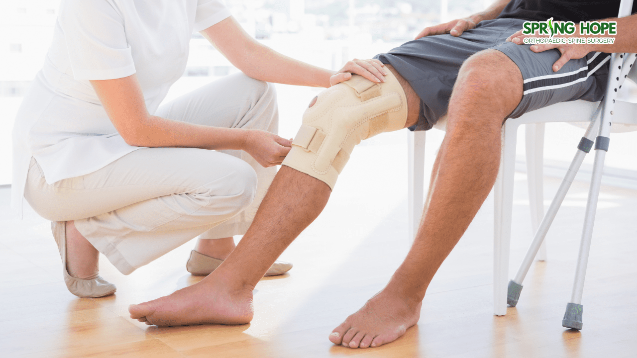
Self-care and Activity Modification Initial management often follows the POLICE principle: Protect, Optimal Loading, Ice, Compression, and Elevation. Protecting the joint with relative rest and using ice packs to manage swelling are fundamental first steps. Crutches or a knee immobiliser may be recommended in the early stages to assist with walking.
Medication Oral anti-inflammatory medicines are frequently used to help control pain and reduce localised swelling during the initial healing phase. These should be used under the guidance of a healthcare professional to ensure they are appropriate for the patient’s overall health.
Physiotherapy and Rehabilitation Physiotherapy is a cornerstone of recovery. A structured rehabilitation programme focuses on:
- Restoring full range of motion through gentle stretching and guided movements.
- Strengthening the quadriceps and surrounding muscles to support the joint.
- Improving balance and neuromuscular control through proprioception exercises.
- Gradual return-to-activity protocols that move from low-impact exercises to sport-specific drills.
Injections Evidence-based injection therapies, such as Platelet Rich Plasma (PRP), may be considered in certain cases. This involves using the patient’s own blood components, specifically growth factors, to facilitate the natural healing cascade within the injured ligament.
Surgical Treatment Surgery is typically reserved for severe cases where the ligament is completely torn or when there is significant rotational instability. It is also considered when the MCL injury is part of a complex multi-ligament injury. Procedures may involve direct repair of the torn fibres or reconstruction using a tissue graft.
Prevention and Lifestyle Management
While not all injuries can be prevented, certain lifestyle considerations may reduce the risk of sustaining a ligament sprain.
Posture and Ergonomics Maintaining proper form and technique during physical activity is crucial. Being mindful of knee alignment during movements like squatting or jumping can prevent unnecessary valgus stress.
Exercise Guidance A proper warm-up programme before engaging in sports is essential to prepare the soft tissues for the demands of the activity. Strengthening the muscles around the knee and hip provides a natural “brace” for the ligaments.
Activity Modification Individuals should avoid pushing past their physical limits and should progress slowly when starting a new sport or increasing training intensity.
Practical Daily Considerations General health factors can influence the body’s ability to maintain and repair connective tissue. Proper nutrition, including adequate Vitamin D, and avoiding habits like smoking—which can delay soft tissue healing—are recommended for long-term musculoskeletal health.
When to Seek Medical Attention
Prompt evaluation is important for ensuring an accurate diagnosis and an appropriate recovery plan. It is advisable to consult a qualified healthcare professional if you experience:
- Persistent symptoms that do not improve with rest.
- Worsening pain or increasing swelling in the knee.
- A sensation of instability where the knee feels like it may buckle or give way.
- Progressive weakness or any numbness in the lower leg.
- Acute symptoms following a significant injury or trauma.
If symptoms persist or worsen, it is advisable to consult a qualified healthcare professional for proper evaluation.
——————————————————————————–
Disclaimer: This page is for general educational purposes only and should not be considered medical advice. Please consult a qualified healthcare professional for personalised assessment and treatment.